Sticky models
History as friction in obstetric education
—
Abstract
Introduction
When it comes to teaching novices the techniques, anatomy, and sheer physics of delivering a baby, teachers face a common conundrum. When it is either too difficult, dangerous, or inappropriate to be involved in actual deliveries, how can educators simulate the slipperiness and mess; the internal movements of the baby that are not visible to the eye; the way a woman’s body changes during labour, with her pain and pushing; or the awkwardness of handling instruments?
As with many such challenges in education, the result is a weird and wonderful array of creative solutions to sharing embodied knowledge. History has a host of examples. In early eighteenth-century Paris, Gregoire, an influential chirurgien-accoucheur (surgeon-midwife), simulated deliveries using a human pelvis, a leather and basketwork uterus, and a real foetus, often in some state of decay. A 1750 commentator saw little educative value in such a device, ‘for let a Part[urient], however difficult, present itself in his Machine, you deliver it as easily as you would turn a cork in a Pail of Water’ (quoted in Owen 2016, 76). Despite two centuries of further practice, Hugo Sellheim’s 1901 adult-size birthing machine was unsuccessful for different reasons. In order to accurately represent the complex movement of the baby through the birth canal, one of Sellheim’s assistants was encouraged to crawl through glass tubes naked while covered in soap (Schlünder 2015). Perhaps unsurprisingly, the assistant got stuck. Apparently, glass tubes cannot adequately reflect a pregnant body, nor can a young obstetrician accurately imitate a foetus.
As these two rather unsuccessful examples illustrate, the synchronicity of movement during childbirth provides a precise degree of friction which is difficult to simulate. Simulations might stick too much, or not enough. In this article, we play with the related concepts of stickiness and friction, which manifest in the material construction of two obstetric simulators located in two medical schools, one in the north of Ghana and one in the south of the Netherlands. Building on Marianne de Laet and Annemarie Mol’s (2000) material-semiotic description of ‘fluid technologies’ – tools lauded for their adaptability, particularly in resource-poor environments – Tom Scott-Smith (2018) has recently defined ‘sticky technologies’ as those tools which offer friction and stability in fast-moving crisis situations. Situating these ideas elsewhere, in the twenty-first-century medical school, for instance, offers an opportunity to complicate the concept and to question the effect of both friction and stability in a given technology.
The past, we argue, is palpably present in the material culture of medicine and medical education. As such, technologies require a degree of translation in order for them to travel effectively across time and space. While the value of sticky technologies derives, in part, from the stability of their actor-networks, the material production of stable biomedical networks has, historically, derived from the Global North and has primarily been made with male physicians in mind. The continued use of these technologies in contemporary contexts has the potential to habitually reify the gendered and imperial histories which are manifest in their material construction. The labour required to translate these stable technologies might also be understood as a form of friction, where past practices and preoccupations ‘stick’ to modern materialities.
These are theoretical issues which, we argue, are best addressed through methodological interventions which bring medical history and ethnography into closer collaboration. To this end, we take seriously the ‘implosion project’, a thought experiment posed by Joe Dumit (2014) after Donna Haraway (1997, 68), which begins with the assumption that ‘any interesting being in technoscience… can – and often should – be teased open to show the sticky economic, technical, political, organic, historical, mythic, and textual threads that make up its tissues’. Pulling at the sticky material threads which are made apparent through ethnographic fieldwork provides a productive framework by which historians and anthropologists can flesh out the ghostly histories which have been seen to ‘haunt’ medical anthropology (Moyer and Nguyen 2018). Working backwards from ethnographic material recorded in fieldnotes and detailed in conversation, the historic threads of present practice are, throughout this paper, imploded in view of oral histories, archival research, and time well-spent in museum collections and hospital storerooms across two continents.
Historians of the body and of biomedicine have, in general, been slow to unify material histories with observations drawn from contemporary contexts (for the historiography of the material history of biomedicine, see: Clever and Ruberg 2014; Schouwenburg 2015; Guerrini 2016). The discursive approach found in many cultural histories of the material world assumes that objects can be read as texts, as vessels of human culture, in order to explain the intellectual environment in which they were constructed and consumed. Science and Technology Studies (STS) scholarship has, by contrast, offered greater agency to the matter surrounding us, showing that culture may be shaped by the material world just as readily as matter is shaped by culture (Haraway 1991; Barad 2007).
Despite its influence elsewhere in the medical humanities, Mol’s (2002) ‘praxiographic’ approach to materiality in biomedicine has rarely been applied to historical contexts (a notable exception is Mak 2012). According to Mol (2002, 32), bodies are not standalone objects but are enacted through practice, drawn out of ‘techniques that make things visible, audible, tangible, knowable’. By co-opting the praxiographic method, histories of medicine can illuminate past practices which have shaped, and continue to shape, both artefacts and their users. In doing so, medical historians can help STS scholars and anthropologists repurpose instruments, tools, and textbooks into agential assemblages which change and fragment both medical practice and medical knowledge. Through our implosion of two obstetric simulators, this paper explores how obstetric simulation can be seen to contribute to the various enactments of parturient and foetal bodies.
As a reflective attempt to recreate bodily function and reproduce embodied knowledge through practice, medical simulation in obstetrics education offers an apposite case study for such historical-ethnographic collaboration. In the use of obstetric simulators, we find the ontologically unstable bodies of medical students engaging with anatomies through agential materialisations of historically specific simulations. As has been shown by Ericka Johnson (2008), junctures in the historical enactment of obstetrics in different parts of the world are reflected in the material construction of obstetric simulation in specific spaces, as well as the uncritical transfer of potentially problematic practices. Exploring a deeper history of materiality, this article highlights how gendered and imperial histories remain present in the construction of contemporary obstetric simulations, as well as in the practices that such simulations promote.
Our study takes place in two medical schools which are tied together by collaboration and a particular focus on clinical skills training, enacted largely through the heavy use of simulation from the first year. The medical faculty at Maastricht University is a relatively new school, established in the south of the Netherlands in the mid-1970s with an initial focus on the training of general practitioners. Maastricht’s medical school is well known in medical education circles as one of the pioneers of the pedagogical theory of Problem-Based Learning (PBL) (Klijn 2016; Servant 2016).[note 1] Founded in the 1990s, the medical faculty at the University of Development Studies (UDS) in Tamale was an attempt by the Ghanaian government to increase the number of doctors practising in the country’s northern savannah, a hinterland which is spatially, socially, and historically distinct from the more affluent south (Bening 2005). Financial assistance from Nuffic, a Dutch NGO specialising in the internationalisation (or Europeanisation) of education, helped shore up the fledging medical school which, with the assistance of Maastricht University and a number of other international collaborators, has been running a full PBL curriculum since the early 2000s. Due to this ongoing collaboration, the syllabi in both schools are similar, but the socio-material conditions – both past and present – are radically different.
Our choice of simulators was determined by what piqued our individual interests and should not necessarily be taken as representative of obstetric simulation in either fieldsite. The sticky threads in these models have stuck with us, as well as with those actively involved in medical education and curricula design. It is in this sense that we expand the concept of stickiness in order to consider how, why, and where these technologies have stuck. In a similar sense, we also consider how the pull of past practice also contributes friction to the continued use of technologies, and how the sticky threads of an imploded material history can affect, direct, or undermine the use of simulation. We argue that the labour required for the translation of material conditions across space and time adds a degree of friction to the ongoing use of such technologies and augments the knowledge which they intend to impart. Practice, as a result, adapts to the material conditions left by past actor-networks. The production of materials encourages the reproduction of practices which, in their cyclical turn, encourages the reproduction of these same materials. This friction is not necessarily problematic, but there is value in its elucidation. In our attempt to do just that, we offer both theoretical questions regarding the role of technology in the reproduction of medical knowledge, as well as a methodological approach to the study of materiality which may offer some preliminary answers.
Maastricht and the knitted uterus
The curricula in both Maastricht and Tamale emphasise the development of clinical skills from an early stage. To this end, both schools have skills laboratories which allow students to practice – on each other and on simulations – the procedures deemed necessary for later clinical work. In the medical school in Maastricht, the ‘Skillslab’ is a standalone building with room enough to be divided up by speciality. Each room is filled with intriguing models and simulations, like toy cupboards for grown-ups wanting to be real doctors. In the obstetrics room there are shelves of labelled leather legless trunks, with hollow spaces and zippered vaginas for cloth babies to squeeze through. The secretaries at the Skillslab care for the models with leather conditioner and hand stitch repairs using leftover surgical twine. There is a large plastic doll lightly coated in years-old glycerine, sticky from many attempted births; and also a plastic placenta, more lifelike to handle than a cloth one, and easier for moving through the models. But for the authors, as for many of the staff, a favourite is the knitted uterus.
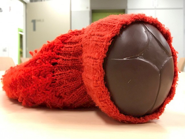
Lying unceremoniously in a drawer in the little room, this is a well-worn and well-loved object. About as long as a forearm, it is knitted in coarse red wool, using a mixture of ribbed and lace stitching. Our second author, Anna Harris, first saw it lying on a tutorial room table, part of the Obstetrics II class set-up: the tools and instruments that the secretaries had arranged for the teacher, to give the lesson about delivering a baby to second year medical students.
The lesson with the knitted uterus goes as follows: a baby doll is inserted into the bulk of the uterus, its head slightly protruding. The teacher then shows how the cervix (i.e., the ribbed cuff) dilates as the head comes through, so that it not only gets wider, but also stretches thin. This helps to show the students something that they cannot see since it is happening inside the body. It is also a way of showing the different positions the baby’s head may take as it descends. While a teacher might talk about the big fontanelles and the small fontanelles (the soft gaps between a baby’s cranial bones) as well as their various positions, this model allows students to see and touch what they should expect to feel. Compared to using diagrams in textbooks to teach these processes, the teachers find using this model to be much more dynamic and simple.
The lesson then becomes more complicated. The teacher takes two blue latex gloves, filling one with a lot of water, and one with less. She then places the full glove over the baby doll’s head, tucking the fingers of the glove into the cuff. A student is asked to feel the bulging glove over the head – this is what intact membranes feel like, they are told. Then the teacher inserts the other glove. Again, a student feels the rubbery bulge – this is what it feels like if the membranes have ruptured higher up, elsewhere in the uterus, but are still intact over the cervix.
After seeing this class for the first time, Anna was captivated by the knitted model, and became even more so as the myths and stories about it unfolded. First, she was told that it was from ‘Africa’, brought back to the laboratory with other models. Yet wool is not often available in African markets, nor is it often employed in African modes of manufacture. Other staff at Maastricht’s medical school explained that it was handmade by a teacher who no longer works at the laboratory, one who had been intimately involved in the internationalisation of the PBL approach employed in Maastricht. Another theory was that it was a ski hat that one of the previous directors thought would make a great teaching tool.
While these mythic histories bear relevance to its current use, the knitted uterus seems to have its conceptual origins in mid-twentieth-century ‘women’s health’ movements, from women’s demands for more medical information about their own bodies, and from the broader societal and medical changes which have been explained in terms of second-wave feminism. It is difficult to say who knitted the first uterus but, in 1940 at least, a knitted uterus formed part of a more extensive, handmade apparatus for the teaching of obstetrics. Time magazine even ran a story on its demonstration, a piece which was largely focussed on the ‘tiny, twinkling, seventy-seven-year-old Dr. Bertha Van Hoosen’ (Time 1940). At a medical conference in Chicago, Dr. Van Hoosen presented a ‘two-foot, mangerlike’ box:
The box represented a woman’s abdomen. Inside, homemade in pink and red, were models of all the organs involved in childbirth. The pelvic cavity was an oval fruit basket. The walls of the box, as well as the pelvis, were covered with pink silk, imitating the peritoneum, glistening lining of the abdomen. Red yarn, knitted by Dr. Van Hoosen herself, showed the pattern of abdominal muscles, Fallopian tubes, ovaries. The mouth of the uterus was knitted in a purl stitch, the body in a plain stitch. Inside the womb was a rubber doll, encased in a bag of Cellophane, attached to the placenta (a dark red knitted cap) by an umbilical cord of red corrugated rubber balloons. (Time 1940)
Time’s rather patronising focus on Dr. Van Hoosen’s gender might be considered routine for a publication of this era. Despite taking place at a general meeting of the American College of Surgeons, this ‘surgical kindergarten, showed scores of women doctors how a Caesarean operation is done’ (Time 1940; emphasis added). Male surgeons would, apparently, not benefit from such a rudimentary elucidation of female anatomy. The Time piece ends with a largely out-of-place vignette about a woman’s gonorrhoea surgery in which Dr. Van Hoosen forced a guilty husband to watch the entire procedure. He apparently fainted, and was revived, three times during its course. The material construction and domestic production of Dr. Van Hoosen’s apparatus is clearly presented as symptomatic of her broader ‘feminine’ approach to doctoring.
The ready dismissal of feminised or domestic forms of material production has not entirely abated in the decades since. A medical educator who helped establish the Skillslab in Maastricht in the 1980s described the controversial introduction of a knitted doll into the paediatric examination skills training. Frustrated at not being able to find a toy doll with flexible enough limbs to demonstrate and practice the hip flexion examination, she headed to the wool shop, bought some cheap yarn, and knitted a series of eight childlike dolls for the Skillslab, filling them with stuffing. The dolls wore striped tops but, most importantly, had legs that could bend easily. The size of the dolls meant the students could cradle them in their hands in order to practice how to fit the baby’s hips and legs into their grasp. The (largely male) body of clinical paediatricians were highly sceptical of this development. Not only were these skills that they thought had to be taught during clinical apprenticeships – not in a ‘laboratory’ outside of the hospital – but they were also being taught with such rudimentary materials. As with the knitted uterus, the material history of this homemade doll moves between the living-room manufacture of those cheap, self-help materials which have been seen to colour the history of women’s health (Murphy 2012) and the teaching rooms of medical schools endowed with creative educators that also know how to knit.
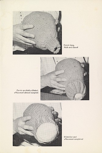
1. The Association of Chartered Physiotherapists in Obstetrics and Gynaecology. Undated. ‘How to make a knitted “uterus” for teaching’. Wellcome Library Archive, SA/CSP/R/2/4/3.
Knitting has, at least during the West’s nineteenth and twentieth centuries, been firmly associated with the feminine. The impermanence and mutability of domestic needlecraft stands in stark contrast with the permanence and public orientation of an idealised masculine production (Goggin and Tobin 2009). Recognising the semiotic effect of such material outputs, the gendering of production has contributed as much to the construction of Western femininity and its attendant politics as the construction of femininity has contributed to the production of a ‘feminine’ material culture (Haraway 1991). As Judith Butler (1993) has prominently argued, the materiality of sex is produced through discursive practice. Others have explained that visual representations of female anatomy played an important role in the discursive production of sexual difference, as well as in the politics of reproduction, and that these historical representations bear relevance for contemporary understandings of the medicalised body (Jordanova 1989; Newman 1996).
Materialisations of female anatomy have long reflected the male gaze of medical practice. Waxwork Venuses – dissected young women reclining on silk, perhaps dressed in pearls, and often in an early stage of pregnancy – are the most obvious example of a male-medico materialisation of a feminine ideal. If, following Butler (1993, 1988), we accept that the materiality of sex is forcibly produced, sustained by force of repetition, and embodied through performance, then embodied understandings of parturient and foetal bodies trace the gendered materiality of the simulations from which they learn. Working from similar ideas, Terri Kapsalis (1997) has questioned whether the growing number of women in medicine will automatically redress the weight of this history. In Maastricht, women now routinely outnumber men in the medical school but, ‘given the implicit hierarchies of the clinic, medical pedagogy, and medical and broader cultural attitudes toward women and women’s bodies, the gynaecological [and obstetric] apparatus helps construct gynaecologists’, even female gynaecologists’, attitudes to women’s bodies’ (Kapsalis 1997, 24–25). While it is impossible to say whether Dr. Van Hoosen saw her knitted uterus as explicitly political, it is a deviation from the gendered materiality of the more traditional obstetric simulations which are described in the second part of this paper. The ideas it embodied offered a reprise from the male-medico performance of obstetric medicine. Indeed, its history in the late twentieth century suggests that the knitted uterus may have endured for precisely these reasons.
It is hard to know where the first uterus was knitted. Perhaps it was Dr. Van Hoosen’s idea, although she makes no mention of it in her discursive biography (Van Hoosen 1948). In any case, its use as a tool for teaching surgeons seems to have fallen away in the years following her 1940 demonstration. Instead, in subsequent decades, the knitted uterus was embraced primarily by antenatal educators, whose foremost interest was the promotion of medicalised childbirth. The concept of ‘maternal impression’, the idea that the development of children in utero were informed by the experiences and actions of pregnant women, was well-developed by the start of the twentieth-century attempts at antenatal education (Al-Gailani 2018). Although direct correlations between maternal morality and foetal monstrosity declined, similar ideas persisted in the science of foetal exposure. By promoting particular forms of behaviour and encouraging the use of hospitals and hospital-trained midwives, early twentieth-century antenatal education was enmeshed in the disciplinary biopower which accompanied Foucauldian explanations of the ‘birth of the clinic’ (Foucault 2003; Al-Gailani 2013). By the mid-twentieth century, however, antenatal education had been repurposed as a biopolitical arm of female emancipation. In 1962, Briefs, the newsletter of New York’s Maternity Center Association, a women’s health education organisation, offered their readership a knitting pattern, a code to the initiated and one with an intimately gendered history. ‘Cast on 48 sts’, begins the pattern. ‘Divide evenly on three needles. Join. K2, P2 until cuff measures 2 inches’. Working through these directions would, apparently, provide a knitted uterus, the primary use of which would be the ‘psychophysical preparation for childbearing’ (Maternity Center Association 1962, 107–8; see also fig. 1).
It was in this later context that the knitted uterus was exported to the Global South. Individuals involved with the United States’ Peace Corps provided instructions for a knitted uterus in a mimeographed book with hand-drawn titles which was first published in 1979. It was well-liked enough to be reprinted in 1985 under the Peace Corps Information and Development Exchange’s ‘appropriate technologies for development’ series (Hansen, King, and Lee 1985). It is worth noting that this tool, which was deemed ‘appropriate for development’ in the 1970s, was also assumed to have contained an appropriate level of technology for an imagined medical school in twenty-first-century Africa. Needlecraft, and especially knitting from patterns, has held a limited role in the gender history of African production. Ironically, in their attempts to emancipate women through antenatal education, the Peace Corps replicated the colonial promotion of needlecraft as an elemental aspect of ‘woman’s work’ (Allman 1994).
In Maastricht, students practice with the knitted uterus because it fills a gap which has still not been addressed by commercially produced obstetric simulators. Its material presence speaks to the sticky histories which have long directed the actor-networks involved in the development of biomedical technologies. At the same time, it is also testament to a longer history of women’s ownership of obstetric technologies. Since the beginning of the twenty-first century, knitting has undergone a significant revival in the Western world. As part of this trend, internet fora propagated a ‘knit-your-own uterus’ movement which originated in a 2004 pattern offered in Knitty, a free online knitting magazine. A year later, the Knit4Choice community encouraged women to drop knitted uteruses on the Supreme Court steps in Washington, D.C., in order to protest the proposed erosion of abortion rights in some states. Unlike Maastricht’s knitted uterus, these are simple, sometimes personified representations of woollen ovaries leading to a woollen uterus. Radical in their softness, they are both a materialisation and a celebration of female anatomy and, as with the Maastricht uterus, have been used as a means to increase individual agency over the politics of reproduction. Some have argued that second-wave feminism involved the denial of traditionally ‘feminine’ crafts in order to break down traditional gender roles, and that the recent revival of knitting has helped to redefine, complicate, and celebrate the history of domesticity and women’s work in the home (Myzelev 2009; Pentney 2008). Yet Maastricht’s knitted uterus suggests a more enduring history. Indeed, both of these knitted patterns share in a gendered performativity and in the soft materialisation of gender politics through knit and purl stitches.
Tamale and the Schultes phantom
In Western imaginations, Africa’s technological landscape is often seen as an absence of technology, or at least an absence of high technology. In what might be considered more generous imaginaries, African technologies are characterised by creative repurposing and reuse, or resourcefulness in resource-poor environments. This is the imagined Africa from which the knitted uterus was thought to have come. In STS, ‘fluidity’ has been used to explore similar elements of Africa’s technological landscape (de Laet and Mol 2000; Redfield 2016). The Zimbabwe Bush Pump ‘B’ was the unlikely hero of de Laet and Mol’s (2000) classic study of ‘fluid technologies’. Locally made and fitting comfortably within a narrative of postcolonial self-definition, the pump’s success derives not from the force of stable networks or powerful inventors but from its simplicity, modesty, and adaptability – all things which seem to have also contributed to the endurance of the knitted uterus.
While not seeking to question de Laet and Mol’s (2000) conclusions, we might consider why fluid technologies have received such attention in subsequent years. Fluid technologies appeal to liberal Western observers sensitive to international imbalances in patterns of consumption, commodity flows, and waste. In this sense, does academic concentration on these forms of technology speak more to predominantly Western cultural backgrounds, anxieties, and presuppositions than to their local value or localised use? Perhaps more importantly, has concentration on fluid technologies overlooked those tools which do fit the bill, the ‘immutable mobiles’ which, in Latour’s (1990) initial explanation of the concept, centred on printed text, the foundational technology of modern education?
Taken as a whole, the technologies in use at UDS’s medical school in the north of Ghana do not comfortably fit either pattern. In the school’s Skillslab, as in Maastricht’s, students learn from the bodies of model patients – sometimes their fellow students, sometimes more literal models. The manikins used for examinations which cannot be conducted on each other (primarily gynaecological, obstetric, and urological exams) are all fairly new, of Western origin, and purchased with two separate donations from UDS’s Dutch partners. Some are highly engineered, like the electronic simulator which allows students to measure foetal heart rates and listen for arrhythmias, but most are not at all mechanical. Budgets and schedules are tight in the medical school; there is little money to develop analogous teaching technologies from local sources and little time to tinker. What has emerged is, therefore, a somewhat unsustainable model for the purchase and maintenance of teaching technologies.
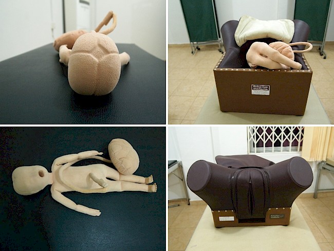
The school’s collection of models and manikins lie in locked cupboards in locked teaching rooms. They are cornerstone technologies of UDS’s Skillslab, and are also rather expensive. In terms of what it helps to teach, if not its material construction, the object most similar to that of Maastricht’s knitted uterus is a large, rubber and leather obstetric simulator, which is produced by the German manufacturer Schultes Medacta and known as the Schultes phantom (see fig. 2). Maastricht has these very same manikins, and uses them in conjunction with other models, such as the knitted uterus, to teach the process of parturition. In fact, Tamale’s first skills coordinator sought the advice of a Maastricht educator involved in internationalisation in order to purchase the most cost-efficient bundle of skills simulators.
Cut off at the top of the torso and the middle of the thigh, a cloth foetus attached to a cloth placenta is inserted into the model’s hollow abdomen and pushed through its stiff, leather vagina in different presentations. The foetus does not pass readily through the model and, by slowing down the lesson, the friction added by the cloth baby and the stiff leather offers students an opportunity to learn. When this simulation is reproduced with a plastic or rubber foetus lubricated with glycerine, as it sometimes is, students are much less able to grasp the mechanics and movements of the child. Though this may be a truer representation of vaginal childbirth (slippery and, in the very final stages, often quick), its educative value lies in the friction granted by its material construction (Scott-Smith 2018).
Unlike the knitted uterus, obstetric phantoms do little to attend to the corporeal changes of the maternal body during labour. These were characteristics which garnered some criticism when the Schultes phantom was first marketed in the nineteenth century; the thick leather was, for instance, unable to accurately represent the way the perineum was stretched by the foetal head during labour (Owen 2016, 148–150). Because of this, other simulators were developed as a necessary addendum to the Schultes models. From these manikins, students primarily learn the various presentations of the foetus, the relative risks to mother and child, and what is required from those attending the birth. Contemporary practice recognises, however, that such manikins offer little attunement to the embodied experience of the patient and that the teacher must ‘perform’ what might be missing, adding personifications to the object for example (Wojcik, forthcoming). In both of our fieldsites, students will stand alongside the phantom, playing the role of the patient. In Maastricht, as in some other medical schools, ‘professional patients’ allow students to examine them, guiding students as to what they feel and how they are made to feel. Other schools employ digital manikins for the same purpose, with pressure sensors built in to show whether the student is palpating the correct, albeit artificial, organ, as well as how much pressure they are exerting on the uterus or the ovaries (Johnson 2008).
2. Schultes Medacta birthing manikin and foetal doll at UDS. Photographs courtesy of Andrea Wojcik.
Operating since 1890, Schultes Medacta is the oldest extant supplier of any medical simulation (Owen 2016, 142–146). The materials which go into the construction of the UDS’s phantom are much the same as those employed by Bernhard Schultze in the nineteenth century. So, by extension, are the philosophies and networks of technology which supported its development. In a period of limited and often dangerous interventions in childbirth, the development of skill with obstetric forceps was an elemental aspect of obstetric education. In a competitive medical market, forceps were initially also a tool for establishing value. In light of this, the first forceps were a family secret, or a succession of family secrets. Likely developed in England around the turn of the seventeenth century, the ‘Roonhuysen’s secret’, as forceps were later known, had been leaked into the Netherlands by the mid-eighteenth century (Drife 2002). We might here assume that the value of forceps, both medically and commercially, derived from an earlier impotence. Disparities in literacy and access to formal education granted men authority over the theoretical, anatomical aspect of obstetric medicine, but the practical side of childbirth, as well as its attendant risks, was still a largely female affair. Male midwives were becoming fashionable in the seventeenth century, in France and later in England, and, with the advent of forceps, men finally had tools to justify their invasion of both the birthing chamber and the body in labour (King 2007; Wilson 1995).
Yet gendered, professional conflict regarding theoretical and practical authority over childbirth cannot, as Margaret Carlyle (2018) has recently argued, explain the development of the obstetric phantom (see also Lieske 2000). Indeed, in eighteenth-century France, women were active in its development and profited from its promotion. Instead, the popularisation of the teaching manikin may be better understood as an outgrowth of the enlightenment promotion of mechanistic approximations of life. Manikins did not, however, develop in isolation. These same enlightenment ideas justified the de-sexing of midwifery – rote interactions with the phantom were hoped to make childbirth ‘mundane rather than titillating’ (Carlyle 2018, 131) – as well as the extension of other technologies into the birthing chamber. Teaching and practicing instrumental deliveries were only made possible with the development of phantoms and, handling nineteenth-century forceps in museum collections today, it is clear that these tools were not made with smaller hands in mind. Obstetric simulators developed in tandem with obstetric forceps, as part of an ecology of technologies and users of technology which was intimately gendered. In response to safer caesarean sections and new imagining technologies, the use of obstetric forceps is of less importance, especially in the Global North, but our heavy leather manikins serve as a resilient reminder of a more complex history than their continued use is wont to acknowledge.
The extension of obstetric technologies across the Global South is a clear example of the biopolitical value of medical materialities in colonial contexts (Vaughan 1991). In imperial Africa, for instance, midwives, obstetricians, and the tools of their trade were usually first used in pursuit of Christian evangelism; state-run hospitals were only established after mission enterprises. In 1911, in what was then the Belgian Congo, one missionary doctor asserted that childbirth was the time of a woman’s ‘greatest spiritual accessibility and receptivity’ and offered an ‘unparalleled opportunity’ to ‘get the girls’ (quoted in Hunt 1999, 201). One of Nancy Rose Hunt’s informants recalled the labour of an anaemic Congolese woman in the 1950s. It was a difficult delivery, but the doctor’s application of forceps helped deliver a healthy baby. ‘The mother, ecstatic about her live baby, wanted to name the newborn after the doctor’s instrument. “Folicepi, Folicepi”, the Congolese mother had chanted’. She was later persuaded to call the baby Emmanuel, ‘for God has been with us in our trouble’ (quoted in Hunt 1999, 196–197).
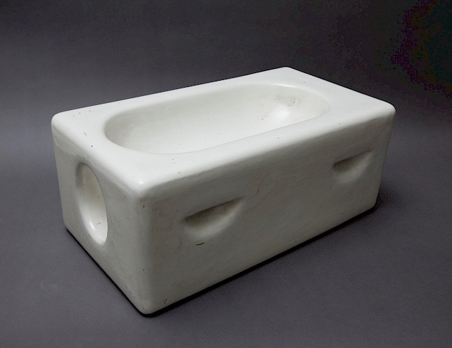
3. A copy of Daniel Dougal’s obstetric simulator. Photo courtesy of New South Wales Ambulance Service Collection, Powerhouse Museum, Sydney. Photographer: Marinco Kojdanovski.
If clinical obstetric technologies offered material justification for European authority over colonised peoples, they were of inherent value to the colonial state. Teaching technologies helped to distribute and mediate African access to these biomedical technologies and attendant practices. Schools training midwives, health attendants, and, later, physicians in the mechanised, European practice of obstetrics emerged in African metropoles from the 1920s. It was in this context that Daniel Dougal, an obstetrician from the University of Manchester, designed a heavy obstetric simulator made almost entirely from white ceramic, with ‘the only extras required [being] a wet towel or thin sheet of sponge rubber to represent the anterior abdominal and uterine walls, and two rubber basins with central openings of different sizes which represent the quarter and fully dilated cervix respectively’ (Dougal 1933, 100–101; see fig. 3). Dougal suggested that cervixes might be made using a rubber ball, rubber draught excluder, and electrical wire. The simulator was heavy, hard-wearing, relatively affordable, easily cleaned, and, materially at least, as far removed from the knitted uterus as possible. Dougal prescribed the explicit use of preserved foetuses, and described their preparation and preservation in much greater detail than the body of the simulator which, by contrast, ‘had the additional advantage that its external appearance in no way resembles the human body and therefore need not be covered or hidden away in a corner when not in use’ (ibid., 100; for a similar device, by a rather remarkable physician, see also Eloesser 1954). Considering the Dougal simulator in light of the knitted uterus, another mid-century obstetric simulator, offers further insight into the gendered materialities which have continued into contemporary medical education. As with Sellheim’s glass birthing machines, the almost exclusive focus on the foetus pares the female body down to a vessel. The rigid construction of the simulator similarly offers some material representation of the fixity of nineteenth-century conceptualisations of sex. If material manifestations of the labouring body can be taken to embody radically different philosophies, then how does the embodied understanding of students learning from material simulations reflect such ideas?
Lamenting, in the 1930s, the expense of available phantoms and ‘the obvious impossibility of providing sufficient clinical material’ (Dougal 1933, 99), the challenges faced by Dougal are similar to those that the UDS medical faculty are currently struggling with. In Tamale Teaching Hospital, where more than four hundred students occupy a six hundred-bed hospital with only twenty to thirty specialist physicians on staff, students on clinical rotation often note that, while they have observed deliveries or attended ward rounds where knowledge of patient health was largely derived from some sensory interaction with pregnant women, they have not necessarily been invited to listen to or feel those same things. Embodied knowledge of obstetrics is, therefore, largely drawn from pre-clinical simulations and usually remains that way until they begin their housemanships. In Tamale, pre-clinical simulation is an absolute necessity. Because of this, greater consideration of the educative technologies is needed. Inattention to the corporeal changes of the maternal body has, however, largely defined the material construction of obstetric simulators. The vast majority of models and manikins from the seventeenth century onwards have taught students to attend to the growth and movement of the foetal body prior to and during labour. Perhaps the knitted uterus is so well liked because it is exceptional in this respect. Perhaps it is exceptional because of its material history; a history which is, ironically, drawn from patient demand for access to their own anatomies. These ideas fit uncomfortably with Madeleine Akrich and Bernike Pasveer’s (2004) suggestion that it was the invention of the ultrasound that changed the nature of the obstetric exam by introducing a more tangible third body into the meeting. Our findings do not necessarily mean to question this, or even the authors’ related conclusion that contemporary obstetrics should not be assumed to form a disciplinary tradition within modern medicine (Akrich and Pasveer 2000). The material history of obstetric simulation does, however, suggest that the erasure of disciplinary practice is complicated by the endurance of historical technologies in the reproduction of medical knowledge.
Offering translation without corruption, obstetric phantoms could be considered standout representations of Latour’s (1990) immutable mobile. They are certainly not fluid. Where de Laet and Mol’s (2000, 225) bush pump ‘doesn’t impose itself but tries to serve’, students in Tamale’s Skillslab serve the obstetric manikins, attending to their histories and their intended publics rather than the histories and publics of northern Ghana. But perhaps the simulators in Tamale are not sticky either, at least not in a material-semiotic sense (Scott-Smith 2018). These are brittle technologies; they look North and are not always able to fully translate. What sticks, then, sticks in the sense of Dumit (2014) and Haraway (1997) – the sticky histories of their construction and the sticking points which constrain and direct their use.
Conclusion
Simulators have long helped medical students, and students of nursing and midwifery, make some sense of the messy, complicated, emotional, and difficult to access process of childbirth. This long history is sewn, or sometimes knitted, into the technologies which students still use to learn obstetrics. The contemporary value and continued use of these simulators require, as a result, a degree of translation. The labour required to translate these technologies across space and time can be understood as attending to friction, the sticking points in the materiality of the object and the practices it promotes. This has the potential to both elevate and to distort the learning experience and the broader value of the technology.
The need to translate technologies is commonly foisted onto people and places with the least agency over the material environment in which they practice and, as such, some technologies promote the uncritical reproduction of practice. The material presence of a homemade, knitted obstetric simulator in Maastricht’s Skillslab speaks to that which, although absent from the mythos surrounding the knitted uterus, remains in its material construction and in the practices which it promotes. The material absence of a similar tool in the locked cupboards at UDS is reflective of a similar material politics but a greater economic burden and longer history of material dependency. Thinking across sites that are materially distinct but which are bound together through materials, concepts, and curricula of modern medical education draws attention to the relevance of the material environment both on the periphery of the actor-network and at its cutting edge.
Attention to these material histories offers insight into the past practices and past lives which have contributed to an imperfect present. It allows their traces to become more visible in ethnographic observations. The particularities of time and place are survived by material legacies and find ways of being replicated, altered, and occasionally even subverted in subsequent practice. Difficult histories of misogyny and imperialism are woven into the fabric of contemporary medicine, and are reified in the continued use of educative simulators in medical schools today. It is only by attending to the cyclical, material reproduction of a problematic past that we might begin to disrupt it.
This is only a first step in the implosion of such technologies, but even our incomplete historical-praxiography of obstetric simulation goes some way to suggest how gendered and imperial histories remain present in the materiality and practice of contemporary medicine. The models we focused on in this article are part of our broader exploration of materiality in medical education across time and space, an exploration which depends on mutual recognition and inspiration between historians and anthropologists. For us, this collaboration offers valuable friction and a chance to see what is sticking – both in the sense of the sticky global threads of history, and the stickiness of using and moving concepts and methodologies across historical and ethnographic investigations to see what shapes and can be shaped by us in our work.
Acknowledgements
This article has been drawn from a larger collaborative project, ‘Making Clinical Sense: A Comparative Study of How Doctors Learn in Digital Times’. For further work from this project, see: www.makingclinicalsense.com. This project receives funding from the European Research Council under the European Union’s Horizon 2020 research and innovation programme (grant agreement no. 678390) for which we are grateful. Our colleagues on this project – Rachel Allison, Carla Greubel, Harro van Lente, Andrea Wojcik, and Sally Wyatt – have contributed significantly to the ideas developed in this article and offered sage advice on earlier drafts. Thanks particularly to Andrea for her generosity in fieldsite introductions and for sharing her thoughts and photos with us – her own work on the role of touch in obstetric simulation is forthcoming. A version of this paper was presented at the Society for the Social Studies of Science meeting in Sydney in 2018; we thank the organisers of the ‘Sensing beyond borders’ panel, Christy Spackman and Nicole Charles, and the attendees for the discussion which emerged. An inspiring writing workshop led by Janelle Taylor helped to flesh out some of the knitted uterus material. Finally, we are especially grateful to our friends in the medical faculties at the University for Development Studies, Maastricht University, and Semmelweis University in Budapest. It is their generosity and openness which has made this research possible.
About the authors
John Nott is a historian and Postdoctoral Researcher in the Maastricht University Science and Technology Studies (MUSTS) research group. His work focuses on the social, economic, and material history of health and wellbeing, primarily in modern Africa.
Anna Harris is an Associate Professor of Anthropology/Science and Technology Studies, also at Maastricht University. Her work concerns issues of learning, sensing (and other bodily practices), and the role of technologies in medicine. She currently leads the project ‘Making Clinical Sense: A Comparative Study of How Doctors Learn in Digital Times’.
References
Akrich, Madeleine, and Bernike Pasveer. 2000. ‘Multiplying Obstetrics: Techniques of Surveillance and Forms of Coordination’. Theoretical Medicine and Bioethics 21 (1): 63–83. https://doi.org/10.1023/A:1009943017769.
Akrich, Madeleine, and Bernike Pasveer. 2004. ‘Embodiment and Disembodiment in Childbirth Narratives’. Body & Society 10 (2–3): 63–84. https://doi.org/10.1177/1357034X04042935.
Al-Gailani, Salim. 2013. ‘Pregnancy, Pathology and Public Morals: Making Antenatal Care in Early Twentieth-Century Edinburgh’. In Western Maternity and Medicine, 1880–1990, edited by Janet Greenlees and Linda Bryder, 31–46. London: Pickering & Chatto. https://doi.org/10.4324/9781315654409.
Al-Gailani, Salim. 2018. ‘“Antenatal Affairs”: Maternal Marking and the Medical Management of Pregnancy in Britain around 1900’. In Imaginationen Des Ungeborenen, edited by Urte Helduser and Burkhard Dohm, 153–172. Heidelberg: Winter-Verlag.
Allman, Jean. 1994. ‘Making Mothers: Missionaries, Medical Officers and Women’s Work in Colonial Asante, 1924-1945’. History Workshop 38: 23–47. https://doi.org/10.1093/hwj/38.1.23.
The Association of Chartered Physiotherapists in Obstetrics and Gynaecology. Undated. ‘How to make a knitted “uterus” for teaching’. Wellcome Library Archive, SA/CSP/R/2/4/3.
Barad, Karen. 2007. Meeting the Universe Halfway: Quantum Physics and the Entanglement of Matter and Meaning. Durham, NC: Duke University Press.
Bening, R. Bagulo. 2005. University for Development Studies in the History of Higher Education in Ghana. Accra: Centre for Savana Art and Civilisation.
Butler, Judith. 1988. ‘Performative Acts and Gender Constitution: An Essay in Phenomenology and Feminist Theory’. Theatre Journal 40 (4): 519–531. https://doi.org/10.2307/3207893.
Butler, Judith. 1993. Bodies That Matter: On the Discursive Limits of ‘Sex’. London: Routledge.
Carlyle, Margaret. 2018. ‘Phantoms in the Classroom: Midwifery Training in Enlightenment Europe’. KNOW: A Journal on the Formation of Knowledge 2 (1): 111–136. https://doi.org/10.1086/696623.
Clever, Iris, and Willemijn Ruberg. 2014. ‘Beyond Cultural History? The Material Turn, Praxiography, and Body History’. Humanities 3 (4): 546–566. https://doi.org/10.3390/h3040546.
de Laet, Marianne, and Annemarie Mol. 2000. ‘The Zimbabwe Bush Pump: Mechanics of a Fluid Technology’. Social Studies of Science 30 (2): 225–263. https://doi.org/10.1177/030631200030002002.
Dougal, Daniel. 1933. ‘The Teaching of Practical Obstetrics’. The Journal of Obstetrics & Gynaecology of the British Empire 40 (1): 99–102. https://doi.org/10.1111/j.1471-0528.1933.tb15525.x.
Drife, James. 2002. ‘The Start of Life: A History of Obstetrics’. Postgraduate Medical Journal 78 (919): 311–315. http://doi.org/10.1136/pmj.78.919.311.
Dumit, Joseph. 2014. ‘Writing the Implosion: Teaching the World One Thing at a Time’. Cultural Anthropology 29 (2): 344–362. https://doi.org/10.14506/ca29.2.09.
Eloesser, Leo. 1954. ‘A Simplified Obstetrical Phantom’. American Journal of Obstetrics & Gynecology 68 (3): 948–949. https://doi.org/10.1016/S0002-9378(16)38341-7.
Foucault, Michel. 2003. The Birth of the Clinic: An Archaeology of Medical Perception. London: Routledge.
Goggin, Maureen Daly, and Beth Fowkes Tobin, eds. 2009. Women and the Material Culture of Needlework and Textiles, 1750-1950. London: Routledge.
Guerrini, Anita. 2016. ‘The Material Turn in the History of Life Science’. Literature Compass 13 (7): 469–480. https://doi.org/10.1111/lic3.12325.
Hansen, Miriam, Karen King, and Barbara Lee. 1985. Preparation for Childbirth: A Health Workers Manual. Peace Corps: Washington, D.C.
Haraway, Donna. 1991. ‘A Cyborg Manifesto: Science, Technology, and Socialist-Feminism in the Late Twentieth Century’. In Simians, Cyborgs and Women: The Reinvention of Nature, 149–181. New York: Routledge. https://doi.org/10.4324/9780203873106.
Haraway, Donna. 1997. Modest_Witness@Second_Millennium.FemaleMan©Meets_OncoMouseTM: Feminism and Technoscience. New York: Routledge.
Hunt, Nancy Rose. 1999. A Colonial Lexicon of Birth Ritual, Medicalization, and Mobility in the Congo. Durham, NC: Duke University Press.
Johnson, Ericka. 2008. ‘Simulating Medical Patients and Practices: Bodies and the Construction of Valid Medical Simulators’. Body & Society 14 (3): 105–128. https://doi.org/10.1177/1357034X08093574.
Jordanova, Ludmilla J. 1989. Sexual Visions: Images of Gender in Science and Medicine between the Eighteenth and Twentieth Centuries. London: Harvester Wheatsheaf.
Kapsalis, Terri. 1997. Public Privates: Performing Gynecology from Both Ends of the Speculum. Durham, NC: Duke University Press.
King, Helen. 2007. Midwifery, Obstetrics and the Rise of Gynaecology: The Uses of a Sixteenth-Century Compendium. Aldershot: Ashgate.
Klijn, Annemieke. 2016. The Maastricht Experiment: On the Challenges Faced by a Young University, 1976–2016. Nijmegen: Vantilt.
Latour, Bruno. 1990. ‘Drawing Things Together’. In Representation in Scientific Practice, edited by M. Lynch and S. Woolgar, 19–68. Cambridge, MA: MIT Press.
Lieske, Pam. 2000. ‘William Smellie’s Use of Obstetrical Machines and the Poor’. Studies in Eighteenth-Century Culture 29: 65–86. https://doi.org/10.1353/sec.2010.0332.
Mak, Geertje. 2012. Doubting Sex: Inscriptions, Bodies and Selves in Nineteenth-Century Hermaphrodite Case Histories. Manchester: Manchester University Press. https://doi.org/10.7228/manchester/9780719086908.001.0001.
Maternity Center Association. 1962. Briefs: Official Publication of the Maternity Center Association. Vol. 26–30. Thorofare, NJ: Slack.
Mol, Annemarie. 2002. The Body Multiple: Ontology in Medical Practice. Durham, NC: Duke University Press.
Moyer, Eileen, and Vinh-Kim Nguyen. 2018. ‘Histories, Hauntings, and Methodological Echoes from Medical Anthropology’s Recent Past’. Medicine Anthropology Theory 5 (1): i–iii. https://doi.org/10.17157/mat.5.1.619.
Murphy, Michelle. 2012. Seizing the Means of Reproduction: Entanglements of Feminism, Health, and Technoscience. Durham, NC: Duke University Press.
Myzelev, Alla. 2009. ‘Whip Your Hobby into Shape: Knitting, Feminism and Construction of Gender’. Textile 7 (2): 148–163. https://doi.org/10.2752/175183509X460065.
Newman, Karen. 1996. Fetal Positions: Individualism, Science, Visuality. Stanford, CA: Stanford University Press.
Owen, Harry. 2016. Simulation in Healthcare Education: An Extensive History. Cham: Springer.
Pentney, Beth Ann. 2008. ‘Feminism, Activism, and Knitting: Are the Fibre Arts a Viable Mode for Feminist Political Action?’. Thirdspace: A Journal of Feminist Theory & Culture 8 (1).
Redfield, Peter. 2016. ‘Fluid Technologies: The Bush Pump, the LifeStraw® and Microworlds of Humanitarian Design’. Social Studies of Science 46 (2): 159–183. https://doi.org/10.1177/0306312715620061.
Schlünder, Martina. 2015. ‘Training the Obstetrical Eye: Teaching Tools for Training the Sense of Touch in Obstetrical Practice (1900-1930)’. Paper presented at Learning How: Training Bodies, Producing Knowledge. Max-Planck Institute for the History of Science, Berlin, Germany.
Scott-Smith, Tom. 2018. ‘Sticky Technologies: Plumpy’nut®, Emergency Feeding and the Viscosity of Humanitarian Design’. Social Studies of Science 48 (1): 3–24. https://doi.org/10.1177/0306312717747418.
Schouwenburg, Hans. 2015. ‘Back to the Future? History, Material Culture and New Materialism’. International Journal for History, Culture and Modernity 3 (2): 59–72. https://doi.org/10.18352/hcm.476.
Servant, Virginie F. C. 2016. ‘Revolutions and Re-Iterations: An Intellectual History of Problem-Based Learning’. PhD dissertation, Erasmus Universiteit Rotterdam.
Time. 1940. ‘Surgery Made Plain’. 4 November.
Van Hoosen, Bertha. 1948. Petticoat Surgeon. London: Peter Davies.
Vaughan, Megan. 1991. Curing Their Ills: Colonial Power and African Illness. Stanford, CA: Stanford University Press.
Wilson, Adrian. 1995. The Making of Man-Midwifery: Childbirth in England, 1660-1770. Cambridge, MA: Harvard University Press.
Wojcik, Andrea. Forthcoming. ‘Training Medical Touch: An Ethnography of Skills Education at a Ghanaian Medical School’. PhD dissertation, Maastricht University.
Endnotes
1 Back
Problem-Based Learning (PBL) involves small tutorial groups, hands-on training, and a limited number of lectures. In PBL-based medical schools, students work in small groups, under the supervision of a tutor, to learn both science and skills from real-life scenarios.


